The Truth Segment: AI diagnoses cardiac fibrosis in minutes

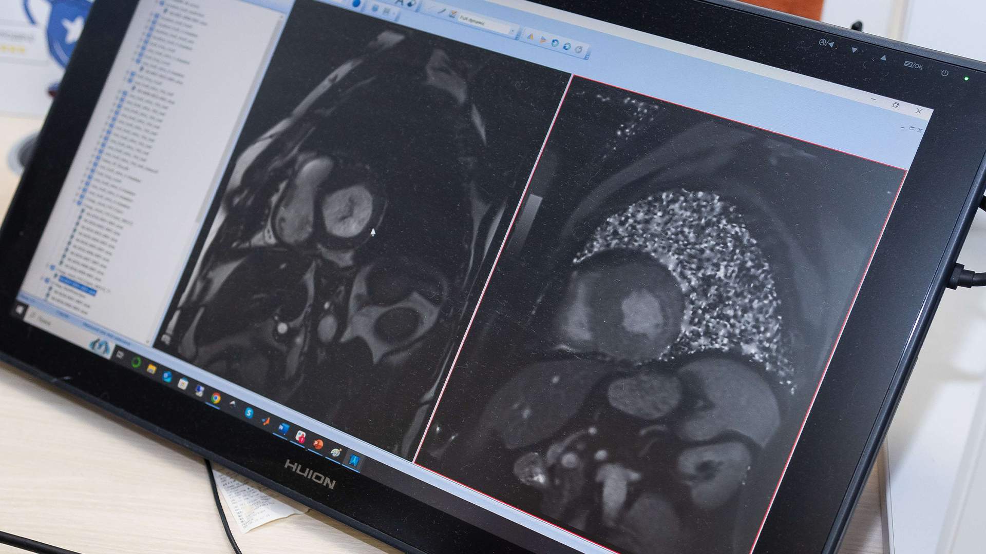
Russian specialists have created an artificial intelligence-based system that can detect cardiac fibrosis by analyzing MRI data. Currently, doctors perform this painstaking work manually, and it can take up to two hours to diagnose one patient. The new technology allows you to do this automatically in just a few minutes. And with the same precision as a human. According to experts, the development will significantly facilitate the work of doctors and speed up the process of choosing a particular treatment, which is very important when it comes to heart diseases.
Neural network for fibrosis diagnosis
ITMO specialists, together with colleagues from the V.A. Almazov National Medical Research Center, have trained artificial intelligence to diagnose cardiac fibrosis using MRI images. To do this, they developed an algorithm that divides the image of the organ into segments, and then determines the location and amount of scar tissue. The development will free doctors from difficult work and accelerate the selection of the most effective strategy for the treatment of heart diseases.
Fibrosis of the heart is an overgrowth of scar tissue that can occur after a myocardial infarction or infectious diseases. One of the methods of its diagnosis is magnetic resonance imaging. However, radiologists have to spend a lot of time on morphometry, that is, accurate measurement of the volume of fibrosis: they manually determine the approximate percentage of fibrous tissue in a particular segment of the heart and enter this information into a table to build a 17-segment diagram. On average, it takes from one to two hours per patient for a doctor to process one series of images. AI will do this in just a few minutes, the developers told Izvestia.
— In the proposed algorithm, the user only needs to mark a few points on the image of the heart and classify the sections, and tissue segmentation and generation of a 17-segment diagram are fully automated. We are currently working on improving our method and developing a faster, fully automatic algorithm that will be able to analyze images instantly without user intervention," said the main
The project's executor, ITMO researcher Walid Al-Haidri.
Attempts to automate image processing using neural networks have been made before. However, the existing models are rather inaccurate and labor—intensive - they require manual or semi-automatic identification of the fibrosis area, that is, the presence of a radiologist. Therefore, the task of automating the generation of 17-segment heart diagrams based on an MRI image remains relevant.
The deep learning model proposed by scientists solves it in stages: first, it determines the area of the heart in which the myocardium is located, then it detects the presence of fibrosis, recognizes the 17 segments into which the heart is divided, and estimates the volume of fibrosis in each of them.
An AI image of the heart in one projection is sufficient for analysis, while doctors may need several images in different projections, which means more time for an MRI scan and its analysis. The neural network was trained on images of the heart, marked up by experts manually, as well as a database of post-infarction MRI scans of this organ. The sample of patients whose images were used was 250 people.
The developers have achieved the accuracy at which the result of the algorithm coincides with the opinions of two experts in 86 and 77% of cases. The authors consider this to be a high indicator: usually the inter-expert agreement is about 80%, that is, the model works almost at the human level.
"We don't just take large datasets and train a neural network to perform routine work on them — we offer doctors a tool that can solve complex tasks at the level of an experienced specialist and will allow them to obtain more information about the relationship between fibrosis localization and other heart parameters," said Ekaterina Bruy, project leader, senior researcher at the ITMO Faculty of Physics.
A quick and objective result of the analysis
Based on data on the location and number of fibrosis, doctors will be able to quickly and accurately predict complications for heart function and disease outcomes, monitor the heart's condition over time, and develop a more effective treatment strategy. In the future, the algorithm can be used not only for processing MRI images, but also adapted for images obtained using computed tomography, the developers are confident.
— Today, artificial intelligence technologies are being actively implemented in various medical fields. One of them is radiation diagnostics, namely the processing of X—ray images, computed tomography and magnetic resonance imaging data. The goal of AI is not only to facilitate the work of a doctor, but also to improve the quality of diagnosis. This is exactly what the development is aimed at. This is certainly important and promising," said Natalia Grigorieva, Director of the Lobachevsky National Research University Institute of Clinical Medicine.
Doctors have a request to reduce data processing time and standardize it. The development is aimed at this, Dmitry Pikhuta, head of the department, radiologist at the RUDN Clinical Diagnostic Center, senior lecturer at the Department of Faculty Surgery at the RUDN Medical Institute, explained to Izvestia.
— The researchers used two methods of segmentation of myocardial fibrosis zones. In the first case, the doctor prepared the study for neural network analysis: all structures not related to the ventricle of the heart were manually removed, the boundaries of the ventricle and its center were determined. The AI analyzed only the presence or absence of fibrosis zones in the presented area. In the second case, all the steps were performed by a set of neural networks. In the first case, the results were more accurate. This suggests that such technology is not yet sufficiently developed to provide accurate data without human intervention," said Dmitry Pikhuta.
However, as the technology develops and improves, it will take its place among the doctor's decision support systems, the specialist added.
According to Dmitry Duplyakov, head of the Department of Propaedeutic Therapy with a course in Cardiology at SamSMU, Chief cardiologist of the Samara region, the proposed system is very useful, as it allows assessing the severity of fibrosis, which directly correlates with the level of heart failure. Thanks to it, it will be easier to predict the patient's condition and its dynamics. The second advantage is speed, and in the case of cardiovascular diseases, this is a critically important factor, he concluded.
Переведено сервисом «Яндекс Переводчик»


