Open the double: future doctors will be taught anatomy on models of their own organs
- Статьи
- Science and technology
- Open the double: future doctors will be taught anatomy on models of their own organs

The Russian Federation is launching the production of personalized, that is, taking into account the characteristics of a particular person, three—dimensional models of bones, joints, muscles, organs, vessels - visual materials that are needed for teaching fundamental disciplines in medicine. Soon, medical students will be able to study anatomy on the "doppelgangers" of their own insides. This teaching method is characterized by an increased interest in the subject and a different quality of training, the authors of the idea said. It will also help to reduce dependence on cadaveric material. Read more about the initiative in the Izvestia article.
Why create copies of the organs of medical students
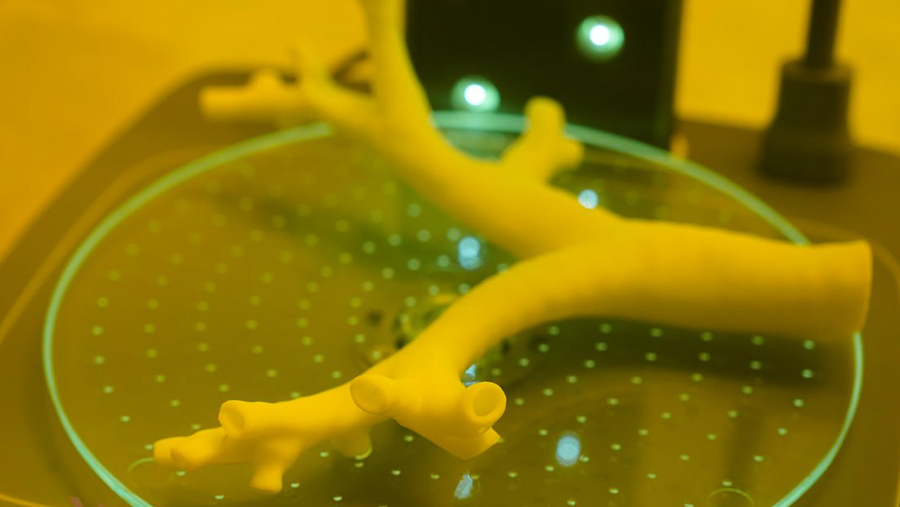
An innovative scientific School of Applied 3D Anatomy has opened at the Department of Human Anatomy and Histology at Sechenov University. It launches the production of three—dimensional models of bones, joints, muscles, organs, vessels - visual materials that are needed for teaching fundamental disciplines in medicine. The printed copies repeat the natural human anatomy with high accuracy and are as detailed as cadaverous (cadaveric) material, the scientists told Izvestia.
According to them, 3D printing provides unlimited variability in the visualization of individual human organs or systems. As a rule, students, residents, and young doctors get used to working with the same simulator and memorize some typical structure. When they encounter an organ with abnormal development, they can hardly deviate from the template. Thanks to three-dimensional printing, you can print as many personalized versions of an organ as you want.
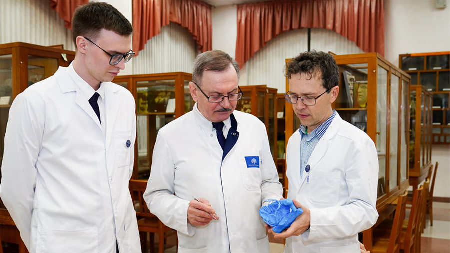
— We can print any organ of people of different ages, with all sorts of morphological parameters, individual variability and various pathologies. Both a student, a candidate for an academic degree, and a practicing doctor can take part in this creative process. We are not far from the fact that students will be able to study fundamental disciplines on their own 3D model," Vadim Kornilov, senior lecturer at the department, told Izvestia.
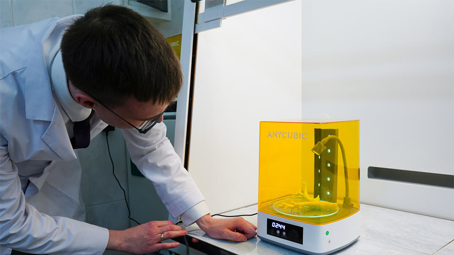
To produce it, you will only need a DICOM file of a computer or magnetic resonance imaging scan of a particular part of the student's body or organ, the teacher specified.
— The teaching methodology, when instead of the "typical human structure" the student studies his own body, is qualitatively different. First of all, an increased interest in the subject and a fundamentally different quality of training. Therefore, we strive to expand this approach as much as possible," said Vadim Kornilov.
At the first stage, the main goal of the engineering school's specialists is to provide high—quality domestic anatomical models from medical universities in the Russian Federation and friendly countries. The technical capabilities of the department make it possible to assemble a "human", including models of organs. But so far we are talking only about small-scale production for our own needs.
— A market analysis is currently underway, and if the high demand for our products is confirmed, the next step will be to attract industrial partners to expand production and the range of 3D models. Combining scientific and technical components, cooperation with industrial partners, as well as the development of a single platform for all simulators will further create a federal-level expertise center for applied 3D anatomy," said Vladimir Nikolenko, Head of the Department.

Application of 3D models of organs
As explained at the university, anatomical simulators and industrial production models are made of plastic by injection molding.
This method of production does not technologically allow recreating many details of anatomical structures, which are clinically important guidelines for the work of future doctors of many specialties.
— 3D printing saves absolutely all the smallest holes and channels, and if something needs to be changed quickly in the model, then they simply make edits to the file, — said Vadim Kornilov.
As for avoiding cadaveric material, as the scientists explained, it will not be possible to completely avoid using cadaveric material in the training of doctors, since a medical professional will treat a person, not a 3D model. But the copies being created will allow for better preparation for practical activities due to the variability of the structure.
Modern 3D anatomical models are certainly a good help for studying anatomy. They provide detailed visualization of complex structures, allow you to practice manual skills and study rare clinical cases. This is a high-tech and effective tool that significantly expands educational opportunities, says Sergey Chemidronov, Head of the Department of Human Anatomy at Samara State Medical University (SamSMU), Associate Professor, market expert at NTI "Helsnet".
— The introduction of anatomical 3D models makes it possible to optimize the use of the traditional cadaver base for solving the most complex educational tasks. At SamSMU, we are actively developing this area, including such technologies in the educational process. But at the same time, cadaveric material retains a fundamental role in medical education, providing tactile authenticity, demonstrating natural anatomical variability and laying the foundations of professional ethics," the expert said.

3D technologies serve as a powerful complement, but they do not replace the unique experience of working with native materials, Sergey Chemidronov summarized.
The use of 3D anatomical models in medical education is an established global practice. Such models are actively used in the training of students, residents and doctors, including for practicing anatomy, surgical approaches and variations of organ structure, commented Albert Rizvanov, head of the Center of Excellence "Personalized Medicine" at Kazan (Volga Region) Federal University, corresponding member of the Academy of Sciences of the Republic of Tatarstan, on the initiative.
— At the same time, many medical universities, including Kazan (Volga Region) Federal University (KFU), have largely abandoned working exclusively with cadaveric material, replacing it with platelets (anatomical objects made by embalming and preserving anatomical preparations. During plastination, water and lipids in biological tissues are replaced by synthetic polymers and resins. — Ed.) and high-precision anatomical modeling. The development of our own 3D anatomy centers and the introduction of such solutions into the educational process certainly deserves a positive assessment," said Albert Rizvanov.
According to the expert, this meets the modern requirements of medical education, reduces dependence on cadaveric material, expands the variability of training and makes it possible to work with individual anatomy and pathologies.
Sechenov University is confident that the opening of the country's first federal Center for expertise in applied 3D anatomy will be possible after the release of anatomical models into mass production.
Переведено сервисом «Яндекс Переводчик»
