Part letter: Data on "extracellular mail" will help better treat inflammation
- Статьи
- Science and technology
- Part letter: Data on "extracellular mail" will help better treat inflammation

Russian scientists were able to "look inside" special extracellular vesicles and learn the features of their structure. This is a kind of molecular "mail" that establishes communication between cells. To do this, the specialists used cryo—electron microscopy, a modern physico-chemical method that allows instant freezing of nanometer-sized samples under conditions close to physiological conditions. As experts told Izvestia, the composition of vesicles changes with inflammation, tumors, neurodegenerative and cardiovascular diseases. Therefore, knowledge of their structure and origin can become the basis for new diagnostic methods and regenerative therapy.
What are extracellular vesicles?
Scientists from the Faculty of Biology and the Faculty of Fundamental Medicine of Moscow State University managed to see the contents of membrane-associated extracellular vesicles (MAV). Extracellular vesicles themselves are membrane—surrounded particles that are released into the extracellular environment. Once in other cells, they are able to trigger serious changes in them. Such structures are a kind of "mail" that establishes communication within the body.
Normally, explosives are present in almost all physiological fluids, they carry complex biochemical signals between cells and take part in almost all physiological processes.
"In many pathologies, the composition of explosives changes, and this can be used to diagnose these diseases," said Anastasia Efimenko, MD, Head of the laboratory at the Center for Regenerative Medicine at the Lomonosov Moscow State University Medical Research and Educational Institute.
According to scientists, an unusual class of membrane—associated extracellular vesicles has recently been discovered among the diversity of explosives. They differ in that they do not travel freely in the medium, but remain attached to the cell membrane from the outside for a long time.
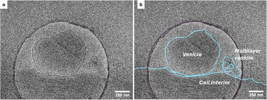
MSU staff conducted explosive research using a cryo-electron microscope. Experts have shown that mAbs are formed from mesenchymal stem cells isolated from human adipose tissue. The lipid membrane surrounding each MAB is perfectly visible when examining frozen samples.
Cryo—electron microscopy is a modern physico-chemical method that allows instant freezing of nanometer-sized samples (macromolecules, viruses, or membrane vesicles) under conditions close to physiological conditions.
Thus, in this study, it was possible to show the presence of multilayer MACS containing several vesicles embedded in each other, surrounded by a lipid membrane. The results obtained will help in studying the physiological role of membrane-associated extracellular vesicles in various processes inside the body.
— The Laboratory of Electron Microscopy of the MSU Faculty of Biology has one of the three cryo-electron microscopes in Russia. Without him, such experiments would not have been possible," said Olga Sokolova, PhD, Professor of Bioengineering at the Moscow State University Faculty of Biology.

The role of extracellular vesicles in inflammation and other pathologies
MSU scientists have managed to literally "look inside life and capture the moment" — to see how explosives look in their natural state, said molecular biologist Arina Kholkina. Using cryo-electron microscopy, they recorded how mesenchymal stem cells secrete membrane-associated vesicles— special particles that do not leave the cell, but remain on its surface.
— Such an observation helps to understand in a new way how cells communicate with each other. It is known that the composition of vesicles changes with inflammation, tumors, neurodegenerative and cardiovascular diseases. Therefore, knowledge of their structure and origin can become the basis for new diagnostic methods and regenerative therapy. MSU's work shows how fundamental research is turning into a tool for medicine of the future," the expert told Izvestia.
For the first time, researchers using cryo-electron microscopy observed multilayer MACS, including several nested vesicles surrounded by a lipid membrane. Such structures can be an important link in intercellular communication — a kind of "nanomesh" between the cell and the external environment, explained Denis Kuzmin, director of the Phystech School of Biological and Medical Physics at MIPT.
— The study of MAB is important from the point of view of understanding the mechanisms of regeneration, inflammation and intercellular signaling. The new data make it possible to revise existing ideas about the ways of communication between cells and lay the foundation for the search for new biomarkers of diseases," the scientist noted.
The project, carried out within the framework of the grant "Molecular technologies of living systems and synthetic Biology", is aimed at a comprehensive study of MAV. The results of the work are published in the journal Microscopy and Microanalysis.
Переведено сервисом «Яндекс Переводчик»
