The doctor spoke about the differences and peculiarities of ultrasound and MRI
Ultrasound (USG) and magnetic resonance imaging (MRI) are the two leading tools that help doctors detect a wide range of pathologies and make informed treatment decisions. Dmitry Ruban, a doctor of ultrasound diagnostics at GASTRO CLINIC, told Izvestia on January 24 how they differ and which method is better suited for each specific case.
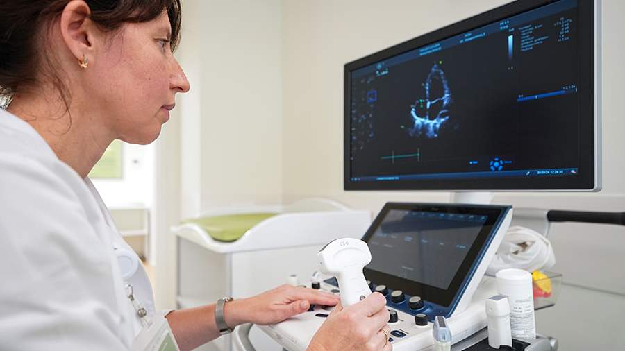
Speaking about the principle of ultrasound, he specified that it is based on the reflection of high-frequency sound waves from tissues. The signals are recorded by a sensor and turned into a real-time image.
MRI uses a powerful magnetic field and radiofrequency pulses. The result is a series of layer-by-layer images that give a detailed three-dimensional view of tissue structure.
"Ultrasound is ideal for rapid assessment of internal organs and soft tissues. Safe in pregnancy, gives the opportunity to study processes in dynamics (heart function, blood flow)," - added the doctor.
At the same time, MRI is preferred for the study of the brain and spinal cord, joints, spine and vessels. It is distinguished by high detail, is able to detect small pathological changes in the early stages.
"Ultrasound does not require radiation exposure, so it is suitable for almost all categories of patients," Ruban specified.
According to him, ultrasound is indispensable in pregnancy and fetal examination. Ultrasound is the main method of monitoring intrauterine development, because it has no harmful effects on the body of the pregnant woman and the child.
In addition, with acute abdominal pain, suspected inflammatory processes (eg, appendicitis) or internal bleeding ultrasound allows you to quickly obtain basic information and guide the choice of further treatment tactics.
At the same time, ultrasound is used for regular monitoring of known pathologies (eg, liver neoplasms) or to study the function of the heart, blood vessels. The procedure can be repeated many times without harm to the patient, said Ruban.

Noting the features of MRI, the expert specified that high image detail is especially useful in complex or doubtful cases, when it is necessary to clarify the nature and localization of the pathological process. MRI of the brain and spinal cord can detect tumors, multiple sclerosis, aneurysms and other pathologies that may remain invisible on ultrasound. In addition, if suspected ligament ruptures, herniated discs, the use of this diagnostic method gives the most detailed picture.
In addition, MRI is carried out in the diagnosis of neoplasms. Thanks to clear tissue contrast, doctors can accurately determine the size, shape and blood supply of the tumor, which is critical when choosing further treatment tactics (surgical removal, radiation therapy, chemotherapy), explained Ruban.
The expert explained that when choosing between ultrasound or MRI, it should be taken into account that in the case of ultrasound quality largely depends on the experience of the doctor and the type of sensor. It is difficult to study organs hidden behind gases (lungs, intestines). And bones for ultrasound are practically "opaque".
In the situation with magnetic resonance imaging, the duration of the procedure is 30-60 minutes, and it is uncomfortable for people with claustrophobia or children. In addition, the doctor noted the high cost compared to ultrasound. Also, if you have metal implants or pacemakers, MRI may be contraindicated.
"Sometimes the choice between ultrasound and MRI is obvious, in other cases it is worth consulting with several specialists. In practice, the doctor is often guided by a combination of factors: where exactly it hurts, how urgently you need to get the result, whether there are contraindications to tomography, whether the patient is ready for a more complex study. The optimal option is often a comprehensive approach: if ultrasound gives insufficient information, the situation is clarified by MRI", - concluded the doctor of ultrasonic diagnostics.
Earlier, December 17, 2024, it was reported that Russian scientists are engaged in the development of the first domestic contrast agent for ultrasound diagnosis. They successfully tested its capabilities in an experiment, obtaining a high-quality image of the heart of a rat. The invention exceeds the characteristics of foreign analogs. This in the future will allow to use it for ultrasound diagnostics in a number of cases where MRI and CT studies were previously used.
Переведено сервисом «Яндекс Переводчик»
